
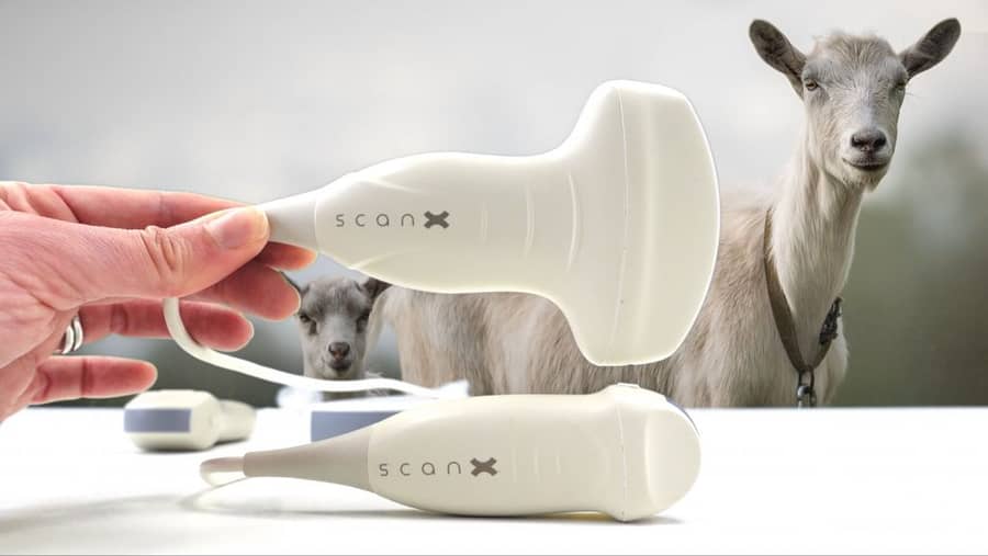
Digital Veterinary X-ray
Fixed and portable digital radiography for veterinary clinics, equine practice and field use across Africa.
Musculoskeletal and thoracic diagnosis in livestock and equines demands clinical-grade image quality. It also has to reach a field without fixed infrastructure.
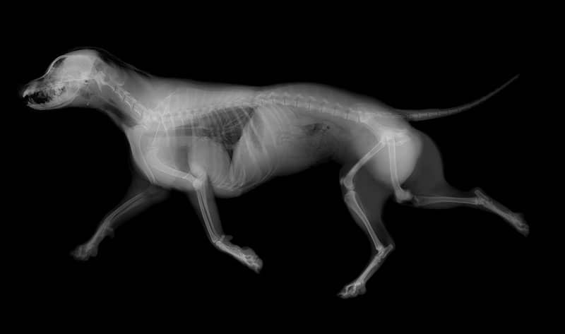
Caldara digital veterinary X-ray systems deliver clinical-grade DR images in fixed clinic and portable field configurations. The flat-panel detector captures high-resolution images of limbs, thorax, abdomen and skull across cattle, horses, sheep, dogs and cats. Images are available in under 10 seconds on a connected workstation or tablet. Jos•Hansen supplies, installs and services Caldara systems with full radiation safety commissioning across East Africa.
No film
Flat-panel digital detectors deliver full-resolution images in under 10 seconds, eliminating film development, chemical processing and darkroom infrastructure. Images are immediately available for reporting and sharing with specialist referral networks.
Two configurations
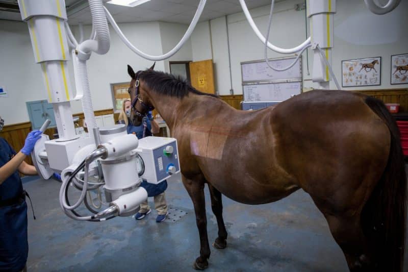
Portable DR systems weigh under 8 kg and run on battery or mains power, deployable in equine yards and on farm visits. Fixed clinic installations use ceiling-mounted or table-column configurations for high-throughput small animal and livestock practices.
Radiation compliance
Jos•Hansen provides full radiation safety commissioning at installation: scatter calculations, personal dosimetry supply, operator training and Radiation Protection Board documentation for Kenya, Uganda and Tanzania.
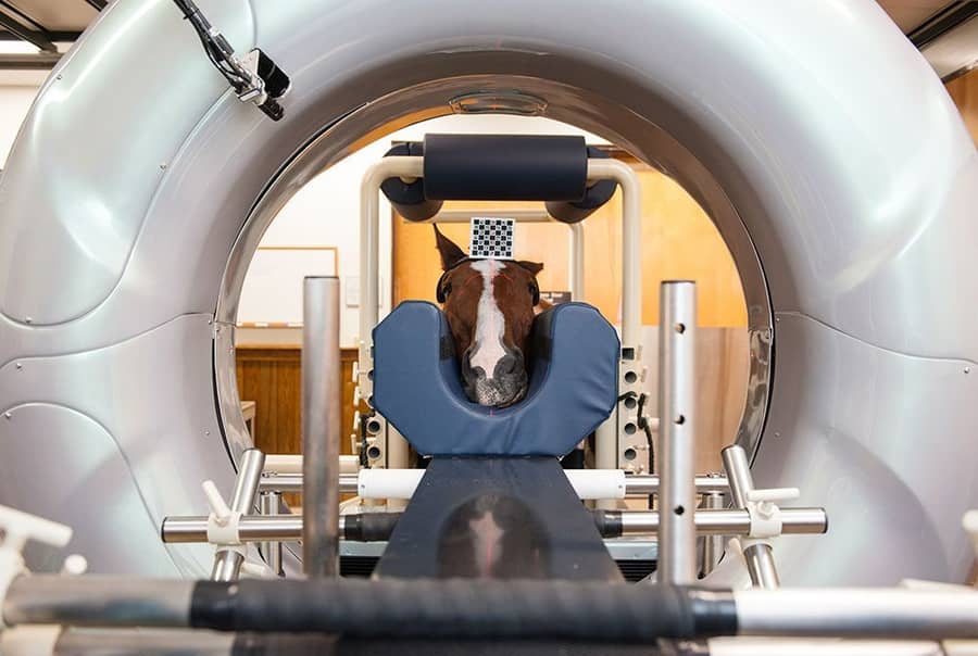
Equine lameness requires 200-micron resolution.
Hoof and fetlock radiographs need to resolve 2 mm joint space changes and subtle pedal bone rotation. Caldara's flat-panel DR delivers 200-micron pixel pitch at the detector, sufficient for reliable laminitis staging, navicular assessment and chip fracture diagnosis in the field without transporting the horse to a referral facility.
Thoracic imaging guides BRD treatment decisions.
Bovine respiratory disease is the leading cause of mortality in feedlot cattle in East Africa. Thoracic radiography differentiates consolidating pneumonia from pleural effusion, abscess and diaphragmatic hernia, changing the treatment decision and the economic outcome per animal. A field-portable X-ray unit pays for itself by preventing a single unnecessary antibiotic course in a high-value heifer.
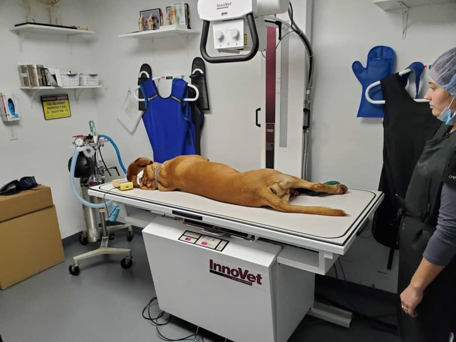
Urban small animal practice demands the same standard.
Companion animal practice in Nairobi, Kampala and Dar es Salaam is growing rapidly. Pet owners expect orthopedic, thoracic and abdominal radiography on demand. Caldara fixed installations deliver the image quality and throughput required for a busy small animal clinic, with DICOM-compliant image storage and telemedicine-ready sharing.
Technical specifications.
Detector type
Flat panel DR, amorphous silicon/cesium iodide
Pixel pitch
200 microns
Image display
Under 10 seconds post-exposure
Generator (portable)
Battery-powered HF, 100mA, 100kV
Configurations
Portable field unit and fixed clinic installation
Software
DICOM 3.0 compliant veterinary PACS
Image available after exposure on flat-panel DR, no film processing required
Pixel pitch delivering fine bone and joint space resolution for equine lameness diagnosis
Standard-compliant image storage and telemedicine-ready sharing for specialist referral
Why Digital.
No film or chemistry
Full digital workflow from exposure to reporting. No consumable film, processing chemicals or darkroom required.
Field portable option
Battery-powered portable unit under 8 kg, deployable for equine field practice and farm visits.
Radiation compliance support
Full RPB commissioning documentation, scatter surveys and operator training at installation.
PACS integration
DICOM-compliant storage and viewer, images accessible remotely for specialist referral and teleconsultation.
Partner with Jos Hansen
Talk to our procurement and operations teams about your next infrastructure, healthcare or scientific deployment.