
Histology & Pathology Systems
Tissue processing, precision microtomy and automated staining for cancer diagnosis.
A cancer diagnosis cannot be made without a pathologist and a slide. The slide quality determines the diagnosis.
Leica Biosystems histopathology equipment enables accurate tissue-based diagnosis from biopsy to stained slide — covering tissue processing, paraffin embedding, precision microtomy and automated haematoxylin and eosin staining. In Africa, where cancer is underdiagnosed because histopathology infrastructure is absent at most hospitals, a functioning Leica workflow at a regional referral centre enables local cancer staging and diagnosis that previously required sample referral abroad. Jos•Hansen installs, trains and maintains Leica Biosystems histopathology platforms across East and Southern Africa — including histotechnician training, reagent supply and preventive maintenance.
Accuracy
Histopathology is the gold standard for cancer diagnosis — the only method that provides definitive tissue classification, grading and staging. A diagnosis made on inadequately prepared slides is a diagnostic risk. Leica Biosystems systems deliver the section quality that pathologists require.
Reproducibility
Automated tissue processing and staining eliminates the batch-to-batch variability of manual methods — producing consistent, diagnosis-ready slides regardless of which technician operated the instrument or which shift processed the specimens.
Completeness
The Leica Biosystems workflow covers every stage from fixation to stained section: ASP300 tissue processor, HistoCore Arcadia embedding centre, RM2255 rotary microtome and ST5020 automated stainer — a complete, integrated histopathology platform from one manufacturer.
The diagnostic foundation of every cancer programme.
Cancer cannot be staged, graded or treated appropriately without a tissue diagnosis. Cytology and imaging guide clinical suspicion — histopathology confirms it. In Africa, where cervical, breast, colorectal and prostate cancers are increasingly prevalent but frequently diagnosed at advanced stage, the absence of local histopathology forces sample referral to national centres with weeks-long turnaround times. A Leica Biosystems workflow installed at a regional referral hospital — and maintained by Jos•Hansen — enables local diagnosis, reduces referral delays and supports the oncology programmes that treatment centres are building.
Automated tissue processing — from biopsy to diagnosis-ready slide.
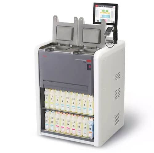
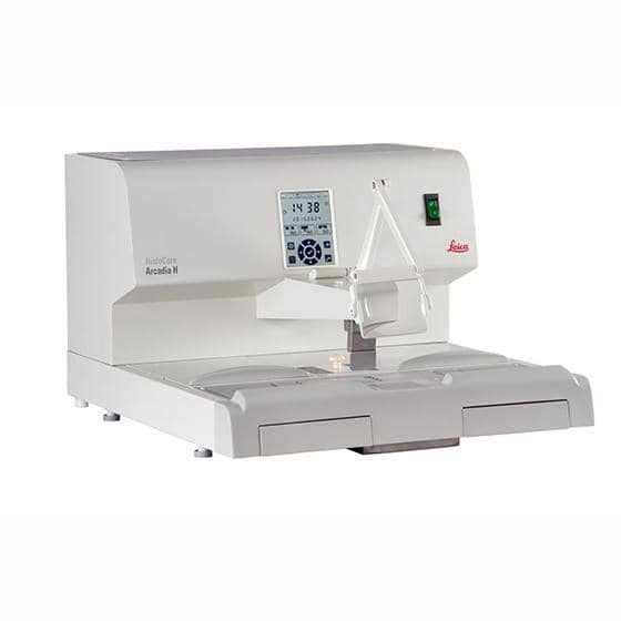
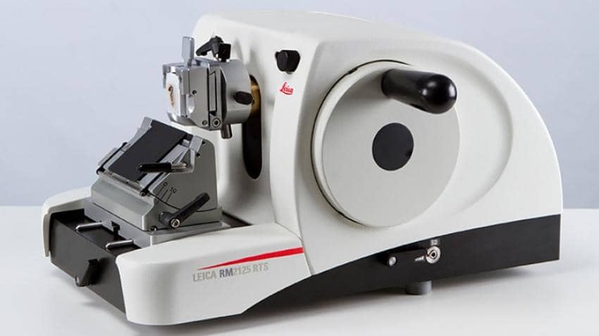
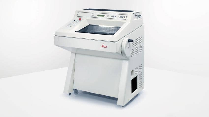
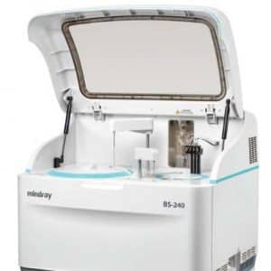
The Leica ASP300 tissue processor runs overnight — dehydrating, clearing and infiltrating biopsy specimens in paraffin through 12 programmable processing stations. The HistoCore Arcadia embedding centre dispenses molten paraffin and positions specimens in orientation moulds for sectioning. The RM2255 rotary microtome sections the embedded block to between 0.5 and 60 μm and transfers sections to the water bath for mounting. The ST5020 automated stainer applies haematoxylin and eosin according to a validated protocol. The result is a stained slide ready for pathologist examination — produced without manual pipetting, timing or technique variation.
Histotechnician training, reagent supply and preventive maintenance — Jos•Hansen delivers.
Leica Biosystems equipment requires skilled histotechnicians to operate it — reagent preparation, embedding technique, section mounting and stain quality assessment are learned skills that determine slide quality. Jos•Hansen provides structured histotechnician training programmes at installation and on an ongoing basis. Reagent supply — xylene, alcohol grades, paraffin, haematoxylin, eosin, mountant and cassettes — is managed through Jos•Hansen's importation and distribution network. Preventive maintenance visits ensure processors, microtomes and stainers remain calibrated and performing to specification.
Technical specifications.
Tissue processor
Leica ASP300 — 300 cassette capacity · 12-station
Microtome
Leica RM2255 — 0.5–60 μm section range
Stainer
Leica ST5020 — automated H&E and special stains
Applications
Cancer · surgical · neuropathology · autopsy
Staining protocols
H&E · PAS · Masson's trichrome · IHC-compatible
Standard
ISO 15189 accreditation support documentation
Minimum microtome section thickness (Leica RM2255) — the precision required for cellular morphology assessment and cancer grading
Cassette capacity per overnight ASP300 tissue processing run — enabling a full day's surgical biopsy workload processed in one unattended cycle
New cancer cases diagnosed annually in Africa — the majority requiring histopathological tissue confirmation for staging and treatment planning
Why Histology.
Gold standard diagnosis
Histopathology is the definitive standard for cancer diagnosis — the tissue section is the only evidence that confirms malignancy, grade and type.
Automated reproducibility
Leica Biosystems automation eliminates manual processing variability — producing consistent diagnosis-quality slides regardless of technician or shift.
ISO 15189 support
Jos•Hansen provides IQ/OQ/PQ validation and quality system documentation supporting ISO 15189 laboratory accreditation for pathology departments.
Complete programme support
Histotechnician training, reagent supply, preventive maintenance and application support from Jos•Hansen across East and Southern Africa.
Partner with Jos Hansen
Talk to our procurement and operations teams about your next infrastructure, healthcare or scientific deployment.